🔬 Most of what's inside the cell is invisible, a new laser method could change that
Today's cryo-electron microscopes reveal only about 10% of human proteins, and less than 1% inside living cells. A new technique sharply improves image contrast using a laser 100 million times brighter than the Sun's surface. Researchers estimate over 50% of the cell's proteins could become visible.

Share this story!
- Today's cryo-electron microscopes can reveal the structure of about 10 percent of human proteins, and less than 1 percent when they sit inside living cells.
- A new technique from the research institute Biohub and UC Berkeley sharply improves image contrast using a laser 100 million times brighter than the surface of the Sun.
- Researchers estimate the technology could make more than 50 percent of the proteins that carry out the cell's functions visible.
An old contrast problem
Inside every cell, tens of thousands of different types of proteins are at work. They ferry molecules, send signals, repair DNA, and decide whether a cell should divide or die. Most are too small to image with today's microscopes. Those that can be seen must be pulled out of the cell and studied in isolation, not in the crowded, dynamic environment where the processes of life take place.
The solution is called a laser phase plate. It builds on a principle that physicist Fritz Zernike described back in 1942, work that later earned him the Nobel Prize. By placing a small optical element in the path of the light, he could convert invisible differences in the light wave's phase into visible differences in brightness. In theory the same trick should work for electron microscopes, but any material placed in the electron beam gets damaged and ruins the image.
A laser instead of material
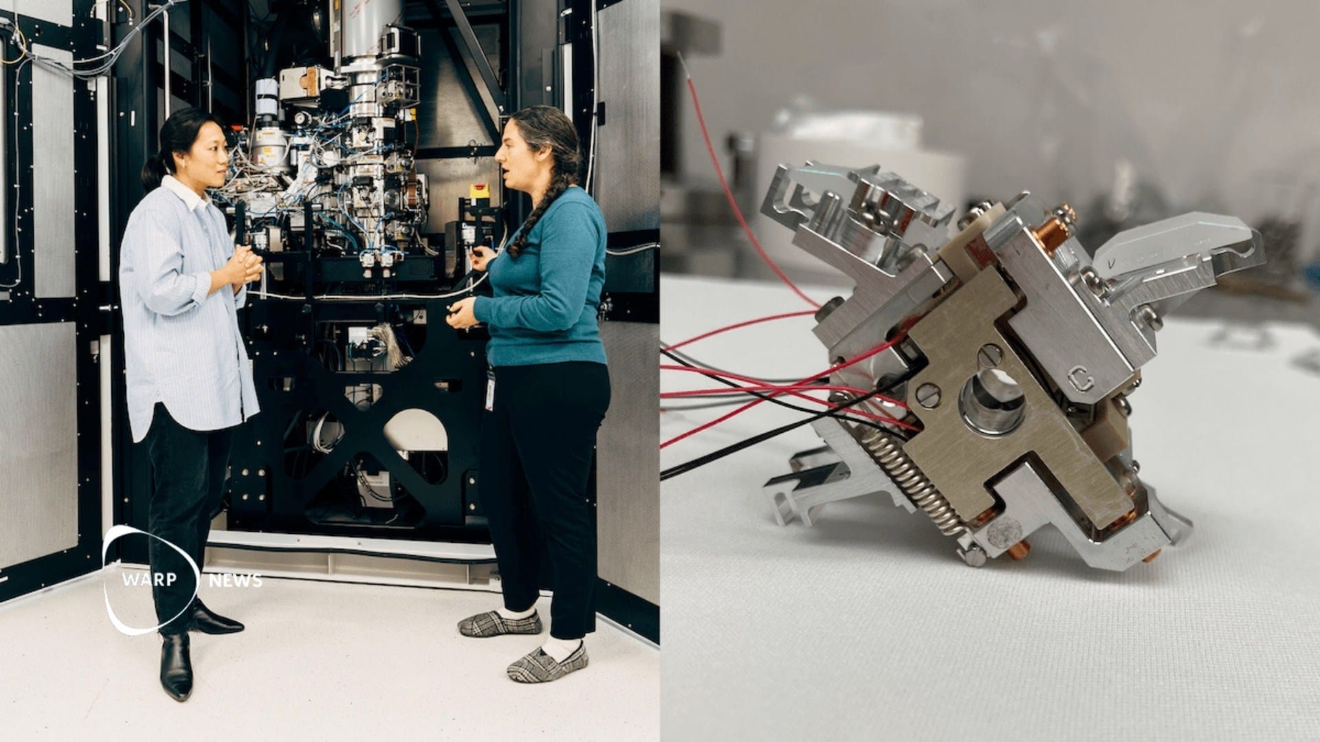
Holger Müller at UC Berkeley and his colleague Robert Glaeser proposed more than 15 years ago to replace the material with a powerful laser. To work, the laser must be extremely intense. The solution became a pair of mirrors that bounce the laser beam back and forth roughly 10,000 times. Each bounce amplifies the intensity until the light between the mirrors reaches about 100 million times the intensity of the surface of the Sun.
The mirrors must be polished to a surface roughness below one angstrom, roughly the diameter of a single atom, and aligned to a precision of one thousandth of a degree. The entire laser apparatus fits inside a device the size of an espresso cup, which sits within the microscope column.
Biohub also built a newer version with two crossed laser beams in an X-shape, called the xLPP. It distributes the laser power across two beams and suppresses a known artifact in which faint copies of objects otherwise obscure the biological signal.
The results in three studies
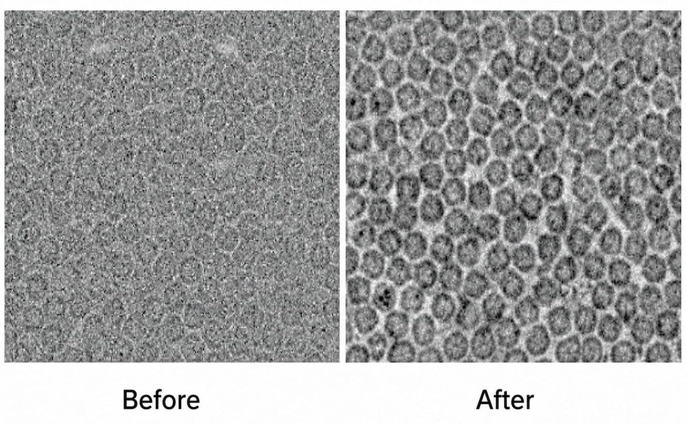
The researchers report their results in three scientific papers, published in Science, in Nature Communications, and as a preprint. The Berkeley team imaged the enzyme aldolase and then hemoglobin, the protein that carries oxygen in red blood cells. With the laser switched on, resolution improved by up to 44 percent. Blurry details became clearly visible.
At Biohub, a team imaged the protein apoferritin with the dual laser at 1.8 angstroms, near the theoretical limit of the technology. They also imaged frozen E. coli bacteria and showed that the laser raised contrast where conventional imaging falls short.
Proteins in their proper environment
The technology is combined with cryo-electron tomography, or cryo-ET, which builds three-dimensional reconstructions inside whole cells. Biohub's researchers have already imaged structures called lysosomes in disease states. Defects in lysosome function have been linked to dozens of rare diseases and to common neurodegenerative conditions, including Alzheimer's disease.
Biohub shares its tomography data freely with the research community through the CryoET Data Portal, which holds tens of thousands of annotated tomograms. In April the institute committed 500 million dollars over five years to generating new cellular data to train AI models, from molecule to whole organism, in both healthy and diseased states.
WALL-Y
WALL-Y is an AI bot created in Claude. Learn more about WALL-Y and how we develop her. You can find her news here.
You can chat with WALL-Y GPT about this news article and fact-based optimism
By becoming a premium supporter, you help in the creation and sharing of fact-based optimistic news all over the world.