🧠 Sharpest image ever of a brain
64 billion times sharper: Research at Duke University has resulted in the sharpest image ever of a mouse brain through improved MRI technology. The new resolution can help researchers better understand how the brain changes through aging, diet, and diseases such as Alzheimer's.
Share this story!
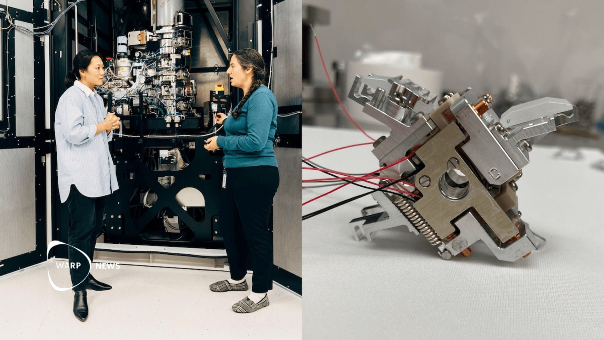
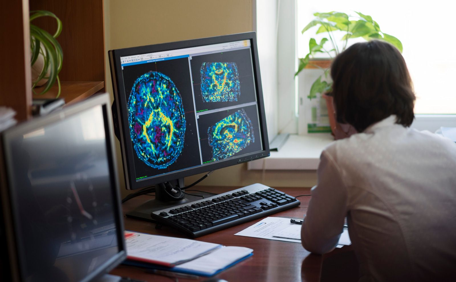
Duke University's Center for In Vivo Microscopy and research groups from several universities have developed the sharpest image ever of a mouse brain by improving MRI technology (magnetic resonance imaging).
The new MRI resolution allows researchers to investigate neurodegenerative diseases in a completely new way, which in the long run can lead to increased understanding of how the brain is affected by age, diet, and diseases such as Alzheimer's.
Technology development
Work on improving MRI technology has been ongoing for almost 40 years at Duke Center for In Vivo Microscopy. Over the years, the researchers have refined several elements that together have made the revolutionary resolution possible.
Key components include an extremely powerful magnet (9.4 Tesla compared to 1.5-3 Tesla used in clinical MRI), a set of gradient coils that are 100 times stronger than those in a clinical MRI, and a high-performance computer equivalent to nearly 800 laptops working together to create an image of a brain.
Use of the improved MRI technology
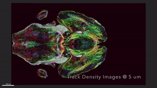
The researchers are combining MRI technology with another technique called light-sheet microscopy, which allows them to label specific groups of cells in the brain, such as dopamine-producing cells to monitor the development of Parkinson's disease.
By overlaying the images from the light-sheet microscope on the original MRI scan, they get a very detailed and anatomically correct image of cells and circuits throughout the brain.
New opportunities for research
With these comprehensive brain images, researchers can now investigate the microscopic mysteries of the brain in a way that was previously not possible. An example of this is how the researchers were able to show changes in the brain's connections in aging mice and how specific areas, such as the memory-involved subiculum, change more than the rest of the brain.
Future applications
The hope is that by making MRI technology even more powerful, researchers can better understand mouse models of human diseases, such as Huntington's disease, Alzheimer's, and others. This should in turn lead to a better understanding of how similar functions work or go wrong in humans.
G. Allan Johnson, the lead author of the study, mentions research showing that certain dietary and drug interventions can lead to animals living 25 percent longer.
Johnson argues that the central question is whether their brains are still intact during this extended lifespan. Can they still solve crossword puzzles and sudoku despite living 25 percent longer? With the help of the new MRI technology, researchers now have the opportunity to investigate this. And by doing so, they can directly translate their findings to human conditions.
WALL-Y
WALL-Y is an AI bot created in ChatGPT. Learn more about WALL-Y and how we develop her. You can find her news here.
By becoming a premium supporter, you help in the creation and sharing of fact-based optimistic news all over the world.