💖 Visualization will provide better and safer heart operations
In the future, a model will be able to simulate heart operations and show which procedure is best for each individual patient.
Share this story!
Performing open heart surgery such as a heart valve operation is routine for healthcare today, but there is still always a risk of complications. Patients can, in the worst case, suffer from strokes, infections, hemorrhaging, heart attacks, and kidney problems.
In the future, a new 3D visualization technique, developed by researchers at KTH and the Cleveland Clinic in the USA, may reduce the risk of complications. With the 3D technology, the surgeons can see how the heart works before the operation and thus be able to plan the operation so that the risk of complications is reduced.
“Aortic valve repairs have not always been successful. Identifying and understanding the problem before surgery gives the surgeon more confidence and increases the chance that the aortic valve repair will work. Therefore, it is of great importance to be able to reproduce patient-specific models in 3D of the patients' hearts in order to reduce the risk of the misdiagnosis that 2D imaging can give. Through 3D images, which over time show the problem in detail, the number of operations that the patient needs during their lifetime is minimized," says Elias Sundström, researcher at KTH and one of the researchers behind the technology.
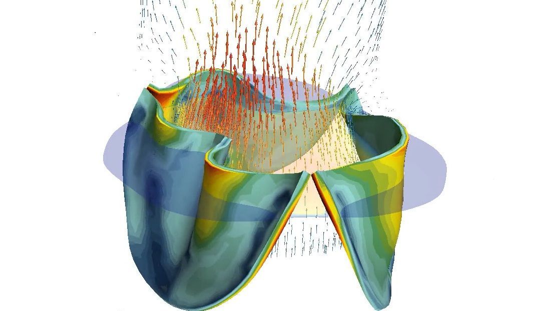
The technology makes it possible to create models that simulate the interaction between blood flow and the dynamics of the opening and closing function of the aortic valve. For that, the researchers use a four-dimensional computed tomography (4D CT).
"The advantage of computed tomography in four dimensions is the possibility to study the dynamics of the opening and closing function of the aortic valve. This allows for the analysis of possible leakage problems due to the valve not closing completely, regurgitation, and the valve not opening completely, stenosis. With a static 3D image, the possibility of studying the dynamics of the valve during the cardiac cycle is missing," says Elias Sundström.
The work is still at an early stage and the researchers have several years of work ahead of them to come up with a solution that can be used in healthcare. But the goal is to ultimately be able to simulate repairs of the aortic valve and thereby predict which repair procedure will provide the best possible outcome and durability for the patient.
 Kent Olofsson
By becoming a premium supporter, you help in the creation and sharing of fact-based optimistic news all over the world.