👂 3D map for the inner ear can provide better treatment of hearing damage
By adapting cochlear implants to each person's cochlea, we can get more effective treatment of severe hearing damage.
Share this story!
A well-functioning cochlea in the inner ear is necessary for us to hear well. Unfortunately, it can also be damaged by loud noises or only weaken with age. Some of the hearing hairs that are in the coil and that register sound are then damaged with the result that we can no longer hear certain sound frequencies.
If the damage becomes severe, a cochlear implant can be inserted to restore some of the hearing. The only problem is that everyone's earbuds look different and this means that not everyone gets the same good effect from an implant.
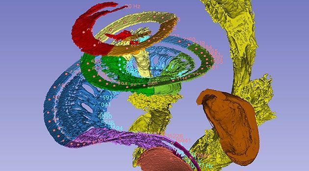
Researchers at Uppsala University and the Canadian universities Western University and the University of Saskatchewan have now developed a method for creating a very detailed 3D map of the cochlea .
The researchers used a synchrotron X-ray, an advanced and powerful form of layer X-ray, to examine the ears. The study was so detailed that the researchers were able to calculate the location of the different frequencies in the cochlea. That information could then be used to create a three-dimensional so-called tonotopic frequency map in the auditory nerve.
- Such a map can be compared to a piano where all frequencies are coded much like the keys. Unlike the piano, which has 88 keys, we have about 3,400 internal hair cells, all of which encode distinct frequencies. The hair cells sit on a 34 millimeter long so-called basilar membrane and are also tuned by 12,000 outer hair cells so that we can hear all the volumes. This information is then passed on to 30,000 nerve fibers that are also tuned for different frequencies, says Helge Rask-Andersen, professor of experimental otology at Uppsala University and one of the researchers behind the study, in a press release.
As mentioned, a cochlear implant can give severely hearing impaired and even deaf back at least part of their hearing . The implant consists of a receiver that is operated into the ear and a processor that is attached to the outside of the skull. The processor receives sounds and converts them into signals sent to the receiver. The receiver in turn converts the signals into electrical impulses that stimulate the auditory nerve.
By getting a better picture of what each person's cochlea looks like, the researchers expect to be able to improve the treatment.
- This can make the treatment with cochlear implants for the hearing impaired more effective, says Helge Rask-Andersen.
Image: Hao Li
By becoming a premium supporter, you help in the creation and sharing of fact-based optimistic news all over the world.